"*" indicates required fields
Types of cataracts
31/08/2017

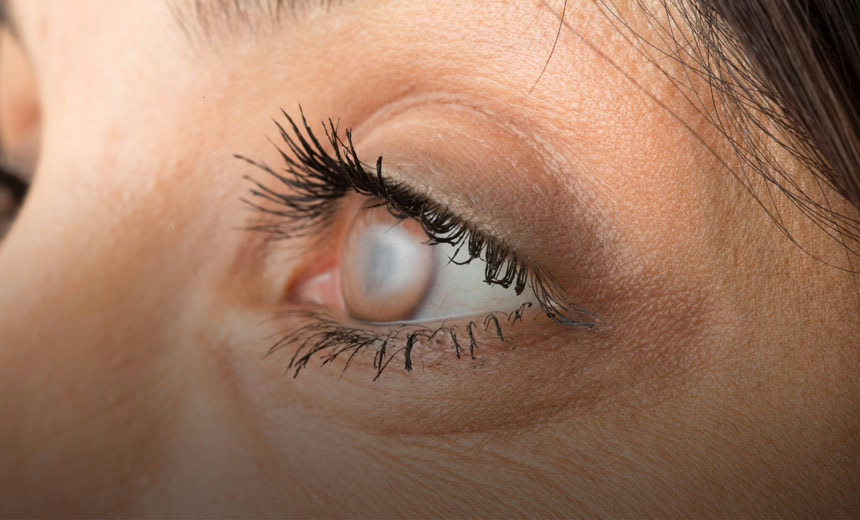
Did you know that there are different types of cataracts?
Cataract surgery is the most performed surgical procedure in the world. However, most people do not realise that there are different types of cataracts.
Ophthalmologists classify cataracts in several ways. The most basic method of classification is according to the location of the actual opacity.
Types of cataracts by location
There are three basic areas in the natural lens of the eye where an opacity can form: the centre portion, the outer part and the back surface.
- Nuclear sclerotic cataract. The most common type of cataract. The centre portion of the natural lens of the eye – the nucleus – becomes yellow and hardens. ‘Sclerosis’ refers to the hardening of the lens nucleus. Deterioration of vision is usually gradual.
- Cortical cataract. A less common form of cataract that appears as a cloudy opacity in the cortex, the outer part of the lens. The cataract resembles spokes that point inwards on the centre of the lens, causing light rays to hit the retina in a scattered fashion. With this type of cataract, the most frequent symptom is experiencing problems with glare.
- Posterior subcapsular cataract. These cataracts form as opacities on the back surface of the lens. They cause light sensitivity, problems with reading vision and glare. They are more prevalent among diabetic patients and those who have taken corticosteroids over a period of time.
The treatment for all three types of cataract is cataract surgery.
Cataract surgery
Today, cataract surgery is considered to be a straightforward procedure. Providing the eye is otherwise healthy, around 98–99% of patients will have a good result.
Cataract surgery involves removing the opacified natural lens and replacing it with an artificial lens called an intraocular lens (IOL). The natural lens is broken up by a process called phacoemulsification, then the pieces are removed via a tiny incision in the side of the cornea (the surface of the eye). The IOL is then inserted through the incision and unfurls into the lens capsule. Sometimes a femtosecond laser is used to complete some of the steps.
Diagnosis of cataracts
If you or someone close to you are experiencing symptoms that indicate the presence of a cataract such as, cloudy or blurry vision, faded colours, glare or poor night vision, book an appointment with a GP or optometrist today.
Read more information about cataracts and cataract surgery
Read the 15 things you need to know about having a cataract operation
References
- The Royal Australian and New Zealand College of Ophthalmologists. Cataract Surgery Online Patient Advisory. Edition 2. Australia: Mi-tec Medical Publishing, 21 February 2019. Available at https://ranzco.edu/policies_and_guideli/cataract/ [Accessed 6 January 2021].
- American Academy of Ophthalmology. What Are Cataracts? USA, 11 December 2020. Available at https://www.aao.org/eye-health/diseases/what-are-cataracts [Accessed 6 January 2021].
- Davis G. The Evolution of Cataract Surgery. Mo Med 2016;113(1):58–62.
- American Academy of Ophthalmology. EyeWiki: Cataract. USA, 30 August 2020. Available at https://eyewiki.aao.org/Cataract [Accessed 6 Jan 2021].
The information on this page is general in nature. All medical and surgical procedures have potential benefits and risks. Consult your ophthalmologist for specific medical advice.
Date last reviewed: 2022-10-05 | Date for next review: 2024-10-05
