Comprehensive eye examination
Your ophthalmologist will take a detailed history, including family history of conditions such as diabetes, high blood pressure or heart disease. You will be asked about any vision problems you are currently experiencing, as well as any that you have had previously. If you use prescription glasses or contact lenses, make sure you take these to your appointment.
The comprehensive eye examination includes tests to determine the health, function and appearance of different parts of the eye.
These may include:
- Visual acuity test to check how well you can see from a set distance using a Snellen chart. This chart contains rows of letters, which decrease in size from the top line to the bottom line.
- Eye muscle test to check the function of the muscles responsible for moving the eye. Your ophthalmologist will hold up and move a pen or object and ask you to follow it with your eyes without moving your neck.
- Refraction test to determine if your vision is normal or if you need corrective lenses. Your ophthalmologist may use a digital refractor or retinoscope to direct a beam of light into your eye and assess how effectively it can focus light. If you have a refractive error, fine adjustments for prescription lenses are made by getting you to look through a phoropter, a mask-like device with multiple lenses which are quickly switched to find out which ones give you the sharpest vision.
- Visual field test to assess your peripheral (side) vision. You may be asked to look into a special instrument and press a button when you see a flashing light. Alternatively, you may be asked to keep your head still, cover one eye and report at what stage you see the ophthalmologist’s moving hand.
- Colour-vision test using multi-coloured dot patterns. Certain colour deficiency will prevent you seeing particular patterns.
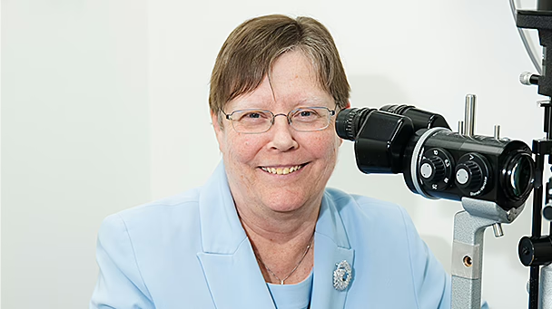
- Slit-lamp examination allows your ophthalmologist to examine the cornea, lens, iris and anterior chamber of the eye. You are asked to sit and rest your chin and forehead on a device that combines a microscope with bright light. Fluorescent eye drops may be used to look for cuts, foreign objects or infections of the cornea.
- Retinal examination to check for diseases of the retina and optic nerve at the back of your eye. After applying eye drops to dilate the pupil, your ophthalmologist uses an ophthalmoscope, slit lamp or bright light mounted on their head to examine each eye. These eye drops cause blurred vision and sensitivity to light for a few hours after the test.
- Tonometry to measure the internal eye pressure, using a device called a tonometer. Eye drops are often used to numb the eye first.
- Pachymetry uses ultrasound waves to measure the thickness of the cornea and look for signs of increased intraocular pressure. Eye drops are used to numb the eye first.
- Neurological exam to check the function of your cranial nerves. This may be required in some situations.
The results of most tests are available straight away, but some may take a few days.
Note: If you have had pupil-dilating drops during the examination, you will have blurry vision and sensitivity to glare and bright light for approximately 4 to 6 hours. You will not be able to drive home and should make other arrangements.
Treatment
If you have a refractive error where the shape of your eyeball prevents light focusing properly (resulting in blurred or distorted vision), your ophthalmologist will prescribe corrective glasses.
If an eye disease is detected or suspected, you may need to have further diagnostic tests before a treatment plan can be recommended. In certain situations, a general ophthalmologist may also refer you to another ophthalmologist who has subspecialised in a particular part of the eye (e.g. a corneal or retinal specialist).
Clinic team
NSW
-


Dr Christopher Go
BMed MD MPH MMed FRANZCO
Locations
- Drummoyne
- Hurstville
Book a consultationwith Dr Christopher Go
Dr Rushmia Karim
BSc MBBS MMed(OphthSc) MMed(ClinEpi)(Hons) Genomics(PGcert) FRCOphth FRANZCO
Locations
- Chatswood
- Drummoyne
- Tuggerah Lakes
Book a consultationwith Dr Rushmia KarimQLD
-
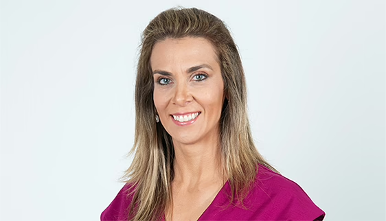
Dr Simone Beheregaray
MD PHD FRANZCO
Locations
- North Adelaide
- Windsor Gardens
- Whyalla
- Brisbane
Book a consultationwith Dr Simone Beheregaray
SA
-

Dr Simone Beheregaray
MD PHD FRANZCO
Locations
- North Adelaide
- Windsor Gardens
- Whyalla
- Brisbane
Book a consultationwith Dr Simone BeheregarayVIC
-

Dr Uday Bhatt
MBBS DTMH DO MSc(EBP) FRCSEd FRCOphth FWCRS FRANZCO
Locations
- Camberwell
- Coburg
- Footscray
Book a consultationwith Dr Uday Bhatt
Dr Jeremy Diamond
MB ChB, PhD, FRCS, FRCOphth, FRANZCO
Locations
- Boronia
Book a consultationwith Dr Jeremy Diamond
Dr Helen Garrott
MBBS(Hons) BMedSc FRANZCO
Locations
- Camberwell
Book a consultationwith Dr Helen Garrott
Dr Philip Hoffman
MBBS(First-class Hons) FRANZCO M Health Ethics Member of ANZSRS
Locations
- Camberwell
- Footscray
Book a consultationwith Dr Philip Hoffman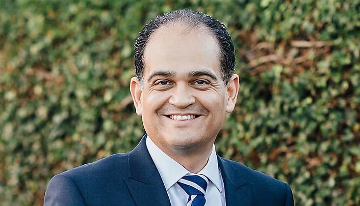
Dr Alex Ioannidis
MBBS FRCOphth FRANZCO
Locations
- Blackburn South
- Camberwell
- Coburg
Book a consultationwith Dr Alex Ioannidis
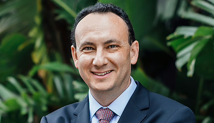
Dr Lewis Levitz
MBBCh MMed FCS(SA)(Ophth) FRCSEd FRANZCO
Locations
- Blackburn South
- Camberwell
- Coburg
Book a consultationwith Dr Lewis Levitz
Dr Nima Pakrou
MBBS(Hons) MMed FRANZCO
Locations
- Footscray
- Boronia
Book a consultationwith Dr Nima Pakrou
Dr Christolyn Raj
MBBS(Hons) MMed MPH FRANZCO
Locations
- Blackburn South
- Camberwell
- Coburg
Book a consultationwith Dr Christolyn Raj
Dr Doug Roydhouse
MBBS(Hons) FRANZCO FRACS
Locations
- Footscray
Book a consultationwith Dr Doug Roydhouse

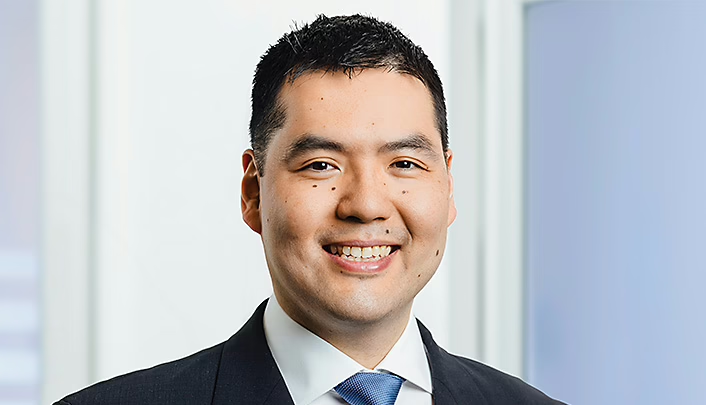
Dr Aaron Yeung
MBBCh, PhD, FRCOphth, FRANZCO
Locations
- Footscray
Book a consultationwith Dr Aaron Yeung
Dr Ian Hurley
MBBS (Melb) FRACS FRANZCO
Locations
- Footscray
- Camberwell
Book a consultationwith Dr Ian Hurley
Dr Daniel McKay
MBBS(Hons) FRANZCO FRCPA
Locations
- Box Hill Retinal Clinic
Book a consultationwith Dr Daniel McKay
Dr James La Nauze AM
MBBS MMedSci(Clin Epi) FRANZCO
Locations
- Footscray
Book a consultationwith Dr James La Nauze AM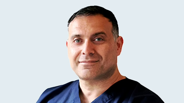
Dr Elvis Ojaimi
MBBS MMed FRANZCO MD
Locations
- Boronia
- Box Hill Retinal Clinic
Book a consultationwith Dr Elvis OjaimiReferences
- Good Vision for Life. 90% of vision loss can be prevented or treated with early detection [Internet]. Australia: Optometry Australia; [date unknown] [cited 2021 Jan 28]. Available from: https://goodvisionforlife.com.au/better-vision/↩︎
ResourcesThe information on this page is general in nature. All medical and surgical procedures have potential benefits and risks. Consult your ophthalmologist for specific medical advice.
Date last reviewed: 2025-02-11 | Date for next review: 2027-02-11


