18/02/2025
Optometrists: This activity may be logged as self-directed learning for 16min of your required CPD hours (dependent upon your personal learning plan).
GPs: This activity may qualify as self-directed learning for 16min of your required CPD hours (educational activities).
Intraocular pressure (IOP) is a particularly useful parameter in evaluating the risk of optic nerve damage. While advances in tonometry technology now allow us to quickly measure a patient’s IOP in the clinic setting, it is just as easy to inadvertently affect the accuracy of the reading. Flow-on effects may include incorrect treatment and management plans and suboptimal patient outcomes, including visual field loss.
Despite the availability of numerous tonometry methods, there is no single, perfect way to measure IOP – each comes with its own pros and cons. Applanation tonometry (based on the Imbert-Fick principle) is most commonly used and assumes a dry, thin-walled, elastic sphere where:
Pressure = Force/Area
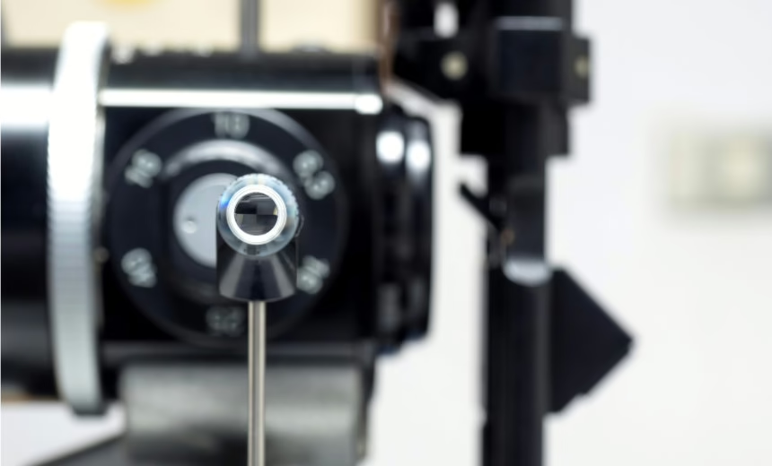
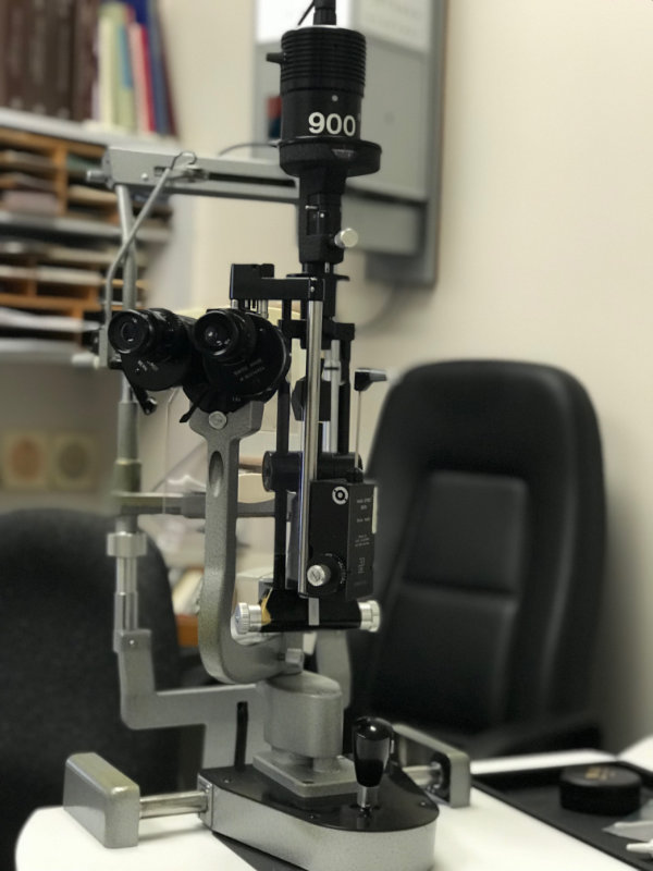
In my opinion, the Goldmann Tonometer (GAT) still remains the gold standard. Introduced in 1954, it measures the force required to flatten a circular corneal area 3.06 mm in diameter (area of 7.35 mm2) – at this size, the amount of fluid displaced is miniscule (around 0.5mL) and the tonometer force becomes equal to the force inside the eye (mmHg).
True, it’s not portable, requires anaesthesia and must be sterilised, but it’s accurate and can detect both lower and higher IOPs (although perhaps not as high as some of the newer devices). Of course, errors can still be made when using GAT to measure IOP – below are some variables to consider.
Of course, there are many other techniques apart from GAT that are useful in providing a quick estimate of IOP. Non-contact ‘air puff’ tonometry is particularly useful in children or anxious adults, although it’s not as accurate as using the GAT.
There’s also been much debate about new technology that allows patients to measure their own IOP. Given that we often rely on single measurements to guide treatment for glaucoma, this would obviously be ideal way to fill in the data gaps. However, a number of key challenges need to be considered:
With careful patient selection and education, there could be an exciting role for self-tonometry in the future.
Dr Doug Roydhouse has considerable experience and expertise in the diagnosis and treatment of a range of ophthalmic conditions, including retinal diseases, cataracts and glaucoma. He currently concentrates his practice on consulting services for patients with general eye conditions. Dr Roydhouse practises at Vision Eye Institute Footscray.
This article is for educational and informational purposes only and may not be directly applicable to your individual patients.
Date last reviewed: 2025-02-18 | Date for next review: 2027-02-18
