
What to do when you suspect a neuro-ophthalmic condition
16/07/2018
CPD hours
Optometrists: This activity may be logged as self-directed learning for 8min of your required CPD hours (dependent upon your personal learning plan).
GPs: This activity may qualify as self-directed learning for 8min of your required CPD hours (educational activities).
A good history is critical
Disorders of the brain and nervous system that affect the eye and vision are often complex to diagnose – most neuro-ophthalmic disorders are not ‘spot’ diagnoses. Consequently, much information can be gleaned when obtaining a thorough from the patient.
While you should have some structure or regularity to the way you take a history, be flexible enough to explore potential flags as you come across them in the course of speaking to the patient.
-
Information about the presenting complaint
- The problem (symptoms)
- The time course*
- The speed of onset and development over time
- Any variability during the day
- ‘Warning signs’ prior to symptom onset
- Any previous episodes (how often and how long)
- Triggering factors
*Remember to distinguish between time of onset vs when the patient first noticed the symptoms (e.g. a patient who rubs one eye when ‘irritated’ and notices that the other eye has no vision will not be able to determine the time of onset).
-
Subsequent questioning
- Pain
- Vision loss
- Diplopia
- Past ocular history
- Glasses or contact lenses
- Eye drops, surgery or laser
- Eye patching or surgery as a child
- Past medical history
- Cancer
- Autoimmune disease
- Diabetes, hypertension, high cholesterol
- Smoking
- Trauma
- Surgery
- Medications (including recreational substances)
- Provides clues to systemic diseases forgotten by the patient
- Can manifest or exacerbate neuro-ophthalmic disorders
- Optic neuropathy – ethambutol, isoniazid, amiodarone, drugs for erectile dysfunction
- Raised intracranial pressure – corticosteroids, oral contraceptive pill, tetracyclines, vitamin A derivatives for acne
- Retinopathy – tamoxifen, hydroxychloroquine
- Double vision – penicillamine, aminoglycoside-induced myasthenia gravis
- Nystagmus – phenytoin, lithium
- Family history of ophthalmic or neurologic disease
- Social history
- Diet/nutrition
Importantly, remember to ask open-ended questions rather than direct questions, as this will provide you with much more detailed answers that can help to guide your subsequent line of questioning.
Ask: ‘how have you been lately?’, ‘any trouble combing your hair?’ or ‘any trouble eating?’
Not: ‘do you have a headache?’ or ‘have you lost weight?’
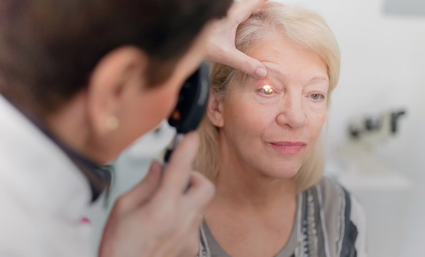
Follow with a thorough clinical examination
When you suspect a neuro-ophthalmic condition, always assess:
- VA
- Colour vision
- VF by confrontation (before dilation)
- Eye movements
- Pupils
- Direct
- Indirect
- Relative afferent pupillary defect (RAPD) to check for optic nerve lesion
- Lid position
- Corneal sensation
- Slit lamp examination (including dilation)
- Optic disc 3Cs – colour, cup, contour
- Take photos
Refer as appropriate
By this stage, you may or may not have a list of differential diagnoses. If referral is appropriate, choose an ophthalmologist with neuro-ophthalmic training to allow further investigations and intervention (if appropriate) in a timely manner.
This article is for educational and informational purposes only and may not be directly applicable to your individual patients.
Date last reviewed: 2023-08-14 | Date for next review: 2025-08-14